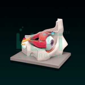
- Mounted on board.
- Shows ventral side, brain stem and spinal cord.
- Branches up to the coccygeal plexus, sympathetic trunk with its connection to the central nervous system.
- Shows 47 positions.
- Made of advanced PVC
A model of the spinal cord in the spinal canal serves educational, medical, and research purposes, providing a detailed representation of the spinal cord within the vertebral column. Here’s a brief overview of its uses:
- Anatomy Education: Used for teaching anatomy, allowing students to study the relationship between the spinal cord and the spinal canal in detail.
- Neurology Training: Beneficial for neurology education, illustrating the spinal cord’s position and protection within the vertebral column.
- Medical Training: Supports medical training programs by providing a tangible model for in-depth study of spinal cord anatomy and its relationship to surrounding structures.
- Orthopedic Studies: Relevant in orthopedic education for understanding the spinal cord’s alignment and protection in the context of musculoskeletal health.
- Neurosurgery Planning: Healthcare professionals may use this model for surgical planning and visualization of procedures related to the spine and spinal cord.
- Patient Education: Enables healthcare practitioners to visually explain spinal conditions, injuries, and treatment options, emphasizing the role of the spinal canal.
- Biomechanics Research: Used in biomechanics research to study the mechanics and movements of the spinal cord within the spinal canal.
- Rehabilitation Training: Practical for training in rehabilitation settings, helping practitioners and patients understand spinal cord injuries and rehabilitation exercises.


