- 30 x 20 x 16cm.
- Made of HSP resin.
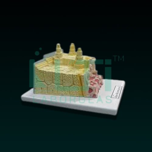
- 3-D section of a lamellar bone.
- It helps to understand the typical elements of a lamellar bone along with the Volkmann & haversian system, spongy & compact parts, endosteum, cortical substance & osteocytes.
- Model shown various planes in cross and longitudinal section.
- 19 features marked.
- Key card/manual provided.
A bone microstructure model serves educational and research purposes, offering a detailed representation of the microscopic structure of bone tissue. Here’s a brief overview of its uses:
- Histology Education: Used for teaching histology, allowing students to study the microscopic features of bone tissue, including osteons, trabeculae, and bone cells.
- Medical Training: Supports medical training programs by providing a close-up view of bone microstructure, enhancing understanding of bone composition and function.
- Orthopedic Studies: Beneficial for orthopedic education, illustrating the intricate details of bone tissue at the microscopic level relevant to musculoskeletal health.
- Biomechanics Research: Used in biomechanics research to study the microstructural aspects of bone that contribute to its mechanical properties and strength.
- Bone Health Education: Aids in educating about bone health by illustrating factors such as bone remodeling, mineralization, and the role of osteocytes.
- Dental Anatomy: Relevant in dental education to showcase the microstructure of jaw bones, aiding in the understanding of dental anatomy and related structures.
- Research Reference: Provides researchers with an accurate model for studying bone microstructure, contributing to advancements in bone biology and related fields.
- Medical Illustration: Useful for medical illustrators to create detailed visuals and representations of bone microstructure for educational materials.

